Rooms 3.11, 3.11.1
Laboratory supervisor: dr Maciej Kania
Laboratory technician: mgr. Adam Wierzbicki
Cathodoluminescence
Cathodoluminescence (CL) is the emission of light from solid matter as a result of being bombarded by a stream of electrons released from a cathode.
Cathodoluminescence (CL) can be used for:
- mineral identification and identification of mineral features (ex. colour, zoning, microfissures, etc.)
- the identification of the progression of diagenetic processes and mineral growth succession in hydrothermal processes
Equipment:
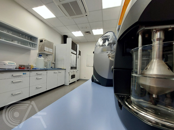
A Cambridge Image Technology Ltd. cold-cathode CLmk3A cathodoluminescence system coupled with a Nikon Eclipse 50T microscope equipped with a Canon EOS 400 or Nikon 5300 camera for image capture (utilizing EOS Utlility software or CoolView software, respectively).
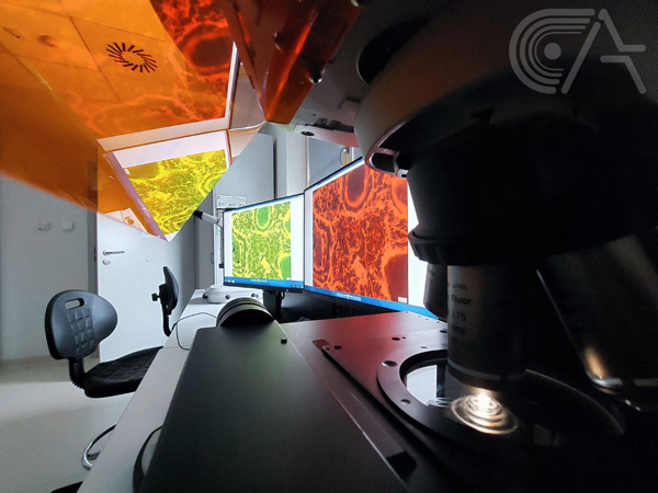
Transmitted light optical microscopy
Analysis of rock and mineral samples using transmitted and polarized light optical microscopy can be used to describe many parameters and features of samples, such as structure and texture as well as the identification of mineral constituents and their form. Such analysis can serve as the basis in deciding which further analyses a sample will undergo, for example, XRD, FTIR, CL or SEM.
Equipment:
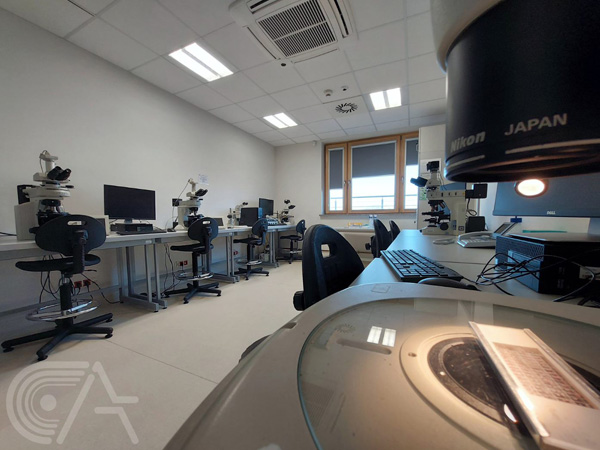
A UV fluorescence optical microscope set (Nikon Eclipse NI polarizing microscope, Prior Lumen 200 UV illumination system, Prior Proscan III stage and controller, Nikon DS. Fi 1C camera, Nikon Digital Sight DS-U3 Camera Control Unit, HP Z440 workstation and NIS-Elements microscope imaging software) - which allows for the automatic scanning of large surface areas of thin sections, horizontal and vertical stacking of images to achieve a 3D-like image, high-resolution image capture and morphometric image analysis.
A Nikon Eclipse E600-POL polarizing microscope equipped with a Canon EOS 400 camera and Canon - EOS Utility software, allowing for image capture.
A Nikon Eclipse E200-POL polarizing microscope equipped with a Nikon Coolpix 950 camera and CoolView software, allowing for image capture.
A Carl Zeiss Jena POL polarizing microscope, which can be equipped with a camera for image capture.
A Carl Zeiss Jena Laboval coupled with an ELTINOR 4 planimetry stage (9 channel, produced by ROW Rathenow, DDR).
Reflected light optical microscopy
Reflected light microscopy allows for the observation of opaque minerals, such as ore minerals and can be used to describe the relationship between ore and gangue minerals. Such analysis can serve as the basis in deciding which further analyses a sample will undergo, for example, XRD, or SEM.
A Nikon Eclipse E600-POL polarizing microscope with an equipped Canon EOS 400 camera and Canon - EOS Utility software, allowing for image capture.
A Carl Zeiss Jena Nephot 221 polarizing microscope, which can be equipped with a camera for image capture.
A Carl Zeiss Jena polarizing microscope, which can be equipped with a camera for image capture.
Rooms -1.03, -1.04
Laboratory supervisor: dr hab. Marta Oszczypko-Clowes
Laboratory technician: dr Piotr Jaglarz
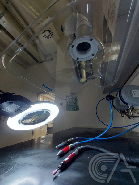
Conducted analyses:
Preparation of polished rock specimens and thin sections of standard thickness for use in polarized optical microscopy. Thin sections are lapped to the desired thickness on a PM5 lapping system and then covered with a cover slip, similarly to biological specimens. However, the use of a cover slip prevents certain further analyses, such as SEM analysis and other chemical analyses. For these types of analyses, lapped thin sections are further polished using a chemically neutral suspension containing diamonds <1 micrometer in width.
Equipment:
- Logitech saws and a Logitech PM5 Precision Lapping and Polishing System with a PLJ2 jig. The PM5 is equipped with a flatness monitor and a series of digital micrometers, which ensure the proper thickness, and flatness of samples. This set allows for the production of 6 standard sized (48x28mm) thin sections at a time.
- A Golz saw
-A Struers Labopol polishing system
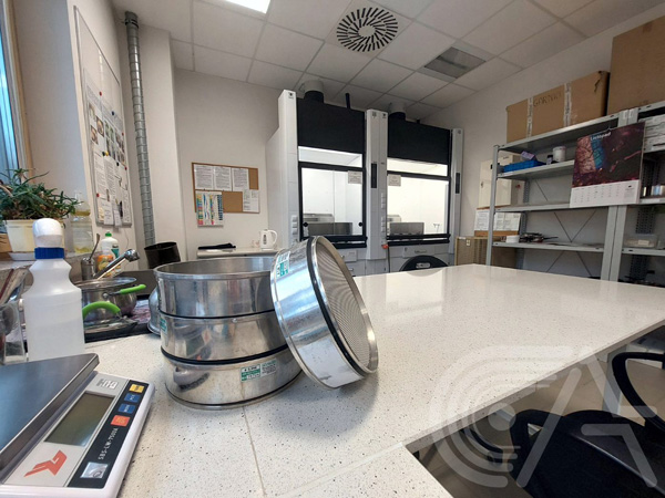
- A vacuum chamber, used for impregnation of porous and loose materials such as soil or sand with resin, prior to processing into thin section.